Radiography
Two forms of radiographic images are in use in medical imaging. Projection radiography and fluoroscopy, with the latter being useful for catheter guidance. These 2D techniques are still in wide use despite the advance of 3D tomography due to the low cost, high resolution, and depending on the application, lower radiation dosages with 2D technique. This imaging modality uses a wide beam of X-rays for image acquisition and is the first imaging technique available in modern medicine.
Fluoroscopy produces real-time images of internal structures of the body in a similar fashion to radiography, but employs a constant input of X-rays, at a lower dose rate. Contrast media, such as barium, iodine, and air are used to visualize internal organs as they work. Fluoroscopy is also used in image-guided procedures when constant feedback during a procedure is required.
Projectional radiographs, more commonly known as X-rays, are often used to determine the type and extent of a fracture as well as for detecting pathological changes in the lungs. With the use of radio-opaque contrast media, such as barium, they can also be used to visualize the structure of the stomach and intestines – this can help diagnose ulcers or certain types of colon cancer.
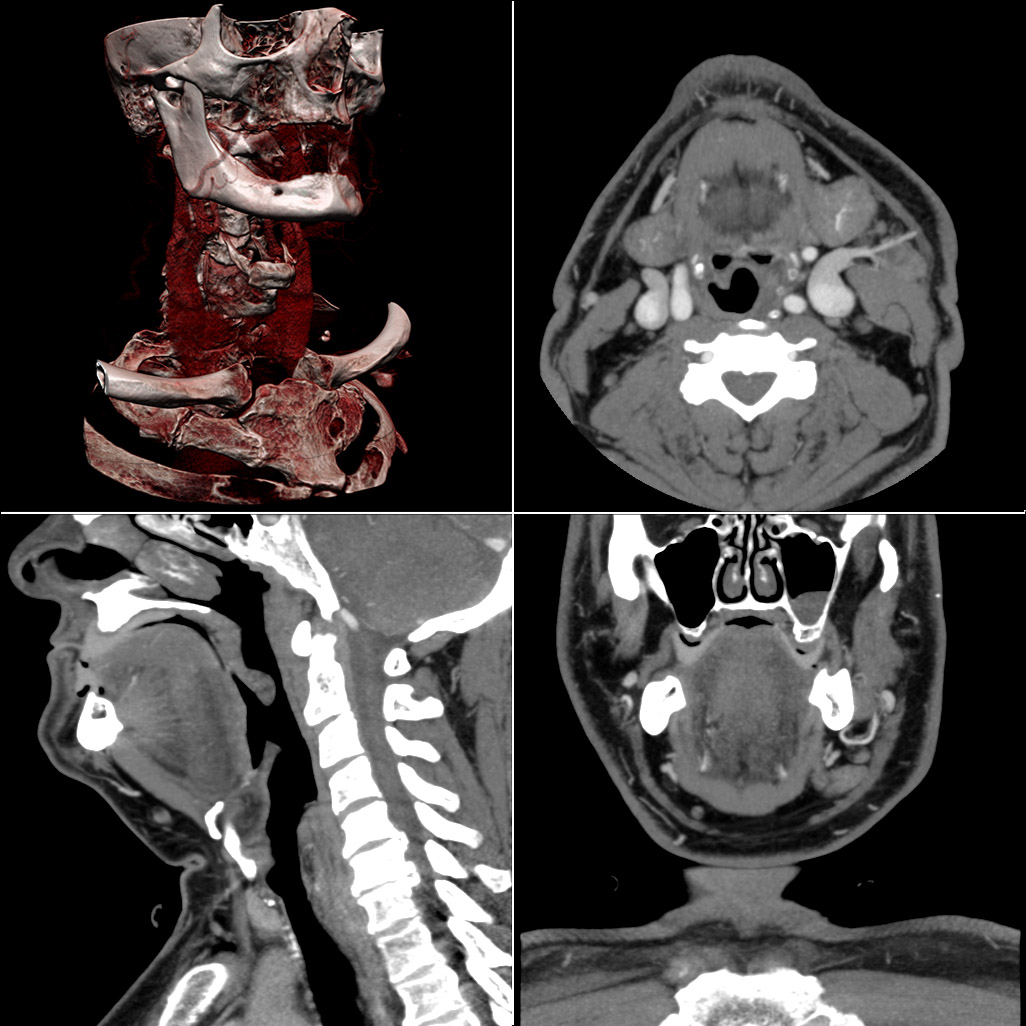
Comprehensive multi-planar reconstruction of neck anatomy for diagnostic assessment.
Magnetic Resonance Imaging
A magnetic resonance imaging instrument (MRI scanner), or "nuclear magnetic resonance (NMR) imaging" scanner as it was originally known, uses powerful magnets to polarize and excite hydrogen nuclei (i.e., single protons) of water molecules in human tissue, producing a detectable signal which is spatially encoded, resulting in images of the body. The MRI machine emits a radio frequency (RF) pulse at the resonant frequency of the hydrogen atoms on water molecules.
Like CT, MRI traditionally creates a two-dimensional image of a thin "slice" of the body and is therefore considered a tomographic imaging technique. Modern MRI instruments are capable of producing images in the form of 3D blocks. Unlike CT, MRI does not involve the use of ionizing radiation and is therefore not associated with the same health hazards.
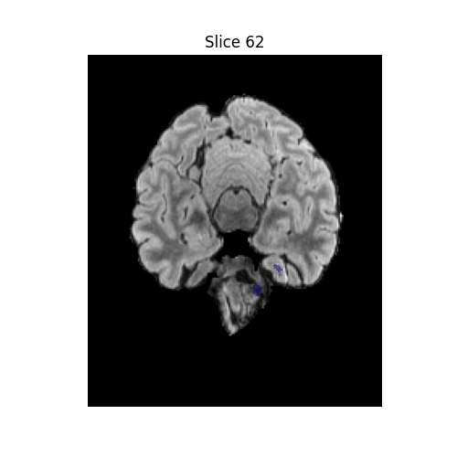
Deep-learning–based extraction of anatomical brain regions for neuroimaging analysis.
Voxel-level tumor delineation from MRI scans for glioma detection and therapeutic planning.
A number of different pulse sequences can be used for specific MRI diagnostic imaging (multiparametric MRI or mpMRI). It is possible to differentiate tissue characteristics by combining two or more of the following imaging sequences: T1-weighted (T1-MRI), T2-weighted (T2-MRI), diffusion weighted imaging (DWI-MRI), dynamic contrast enhancement (DCE-MRI), and spectroscopy (MRI-S). The number of applications of mpMRI for detecting disease in various organs continues to expand, including liver studies, breast tumors, pancreatic tumors, and assessing the effects of vascular disruption agents on cancer tumors.
Ultrasound
Medical ultrasound uses high frequency broadband sound waves in the megahertz range that are reflected by tissue to varying degrees to produce (up to 3D) images. This is commonly associated with imaging the fetus in pregnant women. Uses of ultrasound are much broader, however. Other important uses include imaging the abdominal organs, heart, breast, muscles, tendons, arteries and veins.
Automated segmentation and visualization of pelvic floor musculature for functional assessment.
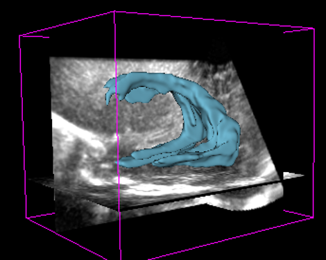
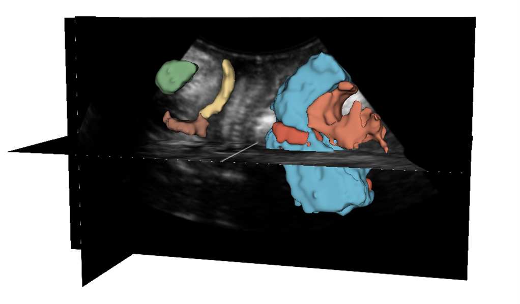
High-resolution 3D rendering of anatomical structures using advanced ultrasound processing pipelines.
While it may provide less anatomical detail than techniques such as CT or MRI, it has several advantages which make it ideal in numerous situations, in particular that it studies the function of moving structures in real-time, emits no ionizing radiation, and contains speckle that can be used in elastography. It is very safe to use and does not appear to cause any adverse effects. It is also relatively inexpensive and quick to perform. Ultrasound scanners can be taken to critically ill patients in intensive care units, avoiding the danger caused while moving the patient to the radiology department. The real-time moving image obtained can be used to guide drainage and biopsy procedures. Doppler capabilities on modern scanners allow the blood flow in arteries and veins to be assessed.